
The Fully Integrated Solution for Automated Analysis of IHC
Why Paige & Mindpeak?
Leverage Paige’s fully open platform to access Mindpeak’s AI applications alongside Paige's robust AI, putting the tools you need for end-to-end diagnosis in one place.
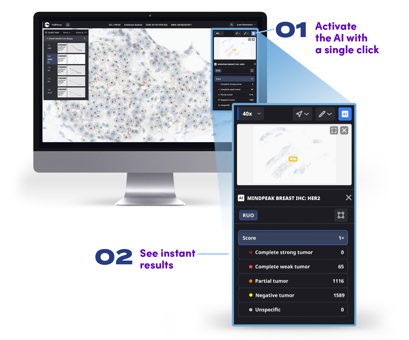
View real-time results from Mindpeak’s IHC biomarker AI algorithms directly within FullFocus®, Paige’s FDA-cleared viewer, creating one seamless workflow to improve lab efficiency.
Reduce the likelihood of error and eliminate tedious manual quantification, simplifying clinical decision-making.
Enhance diagnostic confidence with clinically validated AI support, shown to deliver high performance on images from across different laboratories, stainers, scanners, and antibodies.
A New Approach to IHC Biomarker Quantification
Each Mindpeak algorithm performs automatic tissue segmentation to identify invasive tumor areas and detects and classifies tumor cells to calculate a clinical score per biomarker guideline. The score for the complete tumor on the WSI and the single-cell results are displayed directly in FullFocus for final review by the pathologist.
Mindpeak Breast HER2 | RUO
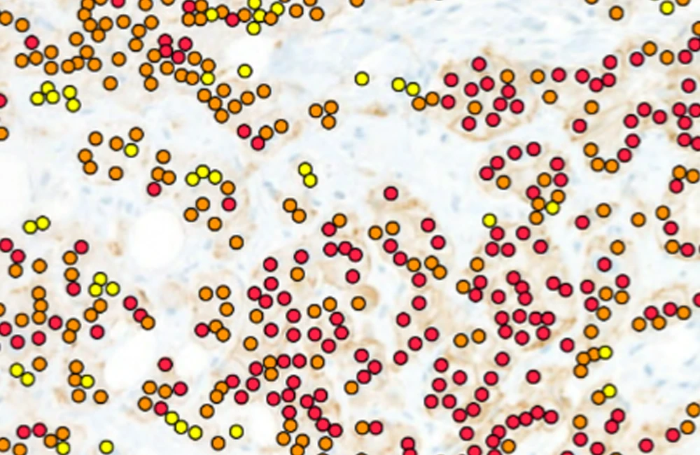
Upon opening an image in FullFocus, both the HER2 score (acc. ASCO/CAP 2018: 0, 1+, 2+, or 3+) for the complete tumor on the WSI and the single-cell results are displayed to the pathologists for final review
Tumor cells are classified as not-stained (negative), partially stained (partial), completely but weakly stained (complete weak), or completely and strongly stained (complete strong), or unspecific
Mindpeak Ki-67 | RUO
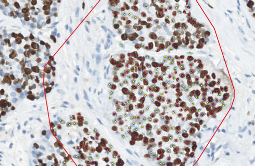
Pathologists can manually select one or many RoIs and the respective proliferation score is directly displayed along with the single-cell results for final review
Hotspot-scoring: An automatic proposal of several pre-scored hotspot alternatives
Global-scoring: The four representative areas according to IKWG-guidelines (negligible, low, medium and strong proliferation) can be automatically proposed, scored and weighted according to overall tumor composition
Mindpeak Breast ER/PR | RUO
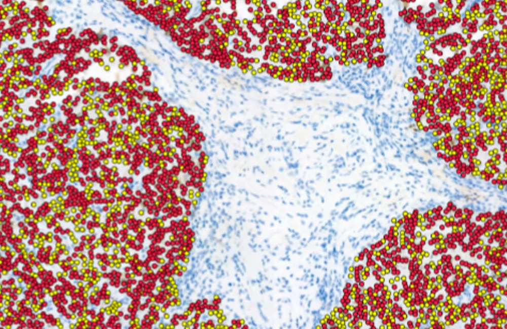
ER and PR scoring is facilitated through automated tissue segmentation in order to identify invasive tumor areas as well as detection, classification and quantification of tumor cells in breast cancer tissue
Upon opening an image in FullFocus both the hormone receptor score (ER or PR) for the complete tumor on the WSI and the single-cell results are displayed to the user for final review
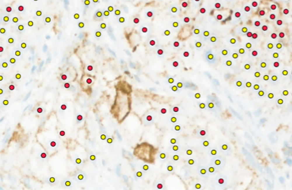
Mindpeak Lung PD-L1 | RUO
PD-L1 scoring is supported through automated tissue segmentation as well as detection, classification, and quantification of tumor cells in non-small cell lung cancer (NSCLC) tissue
Upon opening an image in FullFocus, both the TPS (tumor proportion score) for the complete tumor on the WSI and the single-cell results are displayed to the user for final review